> WOUND HEALING ASSAY
Applications and innovations for 2D in vitro cell migration and invasion studies
July 2024

Applications and innovations for 2D in vitro cell migration and invasion studies
July 2024
We review here the wide range of applications enabled by the in vitro wound healing assay, alongside practical guidelines to study and analyze cell migration. Through quantitative and qualitative analysis, the wound healing assay provides an in vitro method to better understand the movement of cells under well-defined conditions. Processes such as wound healing, angiogenesis, embryogenesis and metastasis can therefore be assessed. Furthermore, the assay allows the analysis of the morphology and polarity of cells during migration. Ultimately, it is an invaluable tool to discover and test new strategies to cure pathologies involving cell migration.
Cell migration is an essential process for the development and maintenance of multicellular organisms [1]. It is involved in many physiological mechanisms such as angiogenesis, embryonic development, immune responses and wound healing. However, when cells develop an invasive behavior, migration can also be involved in pathologies such as cancer metastasis [2]. In vitro studies play a fundamental role in the understanding of cell migration in order to identify new approaches to either enhance or inhibit cell movement. Wound healing assays serve as a standardized approach to mimic cell migration employing a pseudo-wound characterized by a cell-free gap on a cell monolayer. The cells are able to migrate onto the wound area and, therefore, imitate the wound closure as well as other migration processes. The wound healing scratch assay is traditionally the most used technique to create the wound due to its low cost and easy set up. Nevertheless, this method lacks precision and reproducibility [3]. Currently, new ways to perform wound healing in vitro have been developed to enhance consistency in experimental data. Thus, with the use of an in vitro wound healing model, tests and comparisons of the effects of various factors can be achieved while minimizing experimental errors and variations due to the assay itself.
The main applications of the wound healing assay, as documented in research literature, include the study of cell migration (wound healing, angiogenesis, metastasis, embryogenesis), the study of cell morphology, polarity and intracellular organization and drug testing.
Finding novel strategies to stimulate or activate wound healing is necessary to identify solutions to speed up the healing process.
For instance, with the use of a wound healing assay, it was demonstrated that TGF-β loaded exosomes enhance ischemic wound healing [4]. Indeed, the purified exosome product (PEP) delivers TGF-β which promotes the healing process by stimulating cell proliferation and migration.
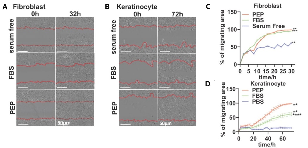
Fig. 1 – PEP accelerates migration into the wound: A wound healing scratch assay reveals that primary rabbit dermal fibroblast (A) and human keratinocytes (B) display wound closure at faster rates when treated with PEP, compared to FBS or PBS. The quantification data (C and D) confirms these results. Image extracted from Shi et al. 2021.
In postnatal life, new blood vessels growth occurs mostly through angiogenesis. This is an important phase in wound healing, supplying oxygen and nutrients necessary for fibroblasts proliferation, collagen synthesis and re-epithelialization [5]. Fibroblast and Endothelial cell migration is essential during angiogenesis [6]. More recent evidence points to the role of macrophage migration for the successful establishment of vascularization [7]. The study of cell migration using the wound healing assay can thus serve to determine the effect of drugs and other factors on angiogenesis.
The impact of exosomes from human umbilical cord blood on wound healing was evaluated by Hu et al. [8]. The experiments performed demonstrated that these exosomes can accelerate cutaneous wound healing via miR-21-3p, which promotes angiogenesis.
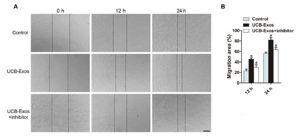
Fig. 2 – MiR-21-3p mediates the pro-angiogenic effects of exosomes (UCB-Exos) on endothelial cells (HMECs). Exosomes induced a significant increase in the motility of HMECs, but the pro-migratory effect was decreased by the miR-21-3p inhibitor, as analyzed by the scratch wound assay (A-B). Image extracted from Hu et al. 2018.
In addition, angiogenesis was studied using Human umbilical vein endothelial cells (HUVECs) [9]. The anti-angiogenic effect of lysyl oxidase propeptide (LOX-PP) in non-transformed endothelial cells migration was demonstrated. Inhibiting angiogenesis with LOX-PP can therefore provide novel therapeutic approaches for treating rheumatoid arthritis, vascular diseases and other pathologies involving excessive angiogenesis.
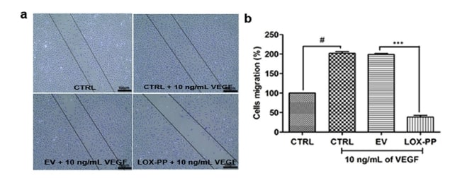
Fig.3 – Lysyl oxidase propeptide negates the pro-angiogenic effect of vascular endothelial growth factor (VEGF) in human endothelial cells. Image extracted from Nareshkumar et al. 2018.
Metastasis is the most lethal manifestation of cancer as it involves the growth of secondary tumors distant from the primary one [10]. The wound healing assay can serve as a valuable tool to better understand the mechanisms involved in tumoral cells migration.
In the study of pancreatic cancer metastasis, a wound healing assay was used as a complementary tool to highlight a regulatory pathway associated with pancreatic cancer progression [11]. Regarding cervical cancer, which has a high mortality rate due to lymph node metastasis, it has been demonstrated that Nrf2 can enhance the migratory capacity of HeLa cells [12]. Identifying approaches to inhibit the expression of Nrf2 may offer a new prospect to prevent or reduce tumoral cell migration and, consequently, avoid, as much as possible, cancer propagation.
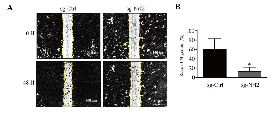
Fig. 4 – Knockout of transcription factor Nrf2 inhibits the migration of HeLa cells. A, B Wound healing assays. Image extracted from Zhang et al. 2023.
Cell migration is an important part of embryogenesis. Ensuring physiological development requires precise cell migration to specific locations during tissue and organ formation [13]. Neural crest cells exhibit migratory behavior within the embryo before differentiating into different cell types that play a crucial role in the development of cartilage, bone, muscle and connective tissues for the skull and facial structures [14]. Abnormalities in the migration of neural crest cells during embryogenesis can lead to cleft lip and or palate, which is considered the most typical craniofacial malformation. An experiment using wound healing assay demonstrated the combined effect of E-cadherin knockout and TNFα to slow down neural crest cells migration [15]. The requirement of cadherin on neural crest migration was thereby confirmed as well as the effects of inflammation on craniofacial development by neural crest cell migration.
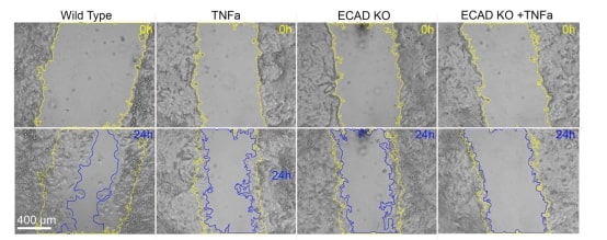
Fig.5 – Combined effect of E-cadherin loss-of-function and pro-inflammatory activation supports a 2-hit model for E-cadherin loss in the neural crest. Scratch assays using hiNCCs in wild type, TNFα exposure, heterozygous E-cadherin knockout (ECAD KO) and combined ECAD KO + TNFα conditions evidencing scratch areas at 0h (yellow line) and 24h (blue line) after scratch in each condition. Image extracted from Avizi et al. 2023.
Prior to migration into the wound, cells reorganize their internal structure, a process known as polarization, in order to reorient themselves towards the wound and effectively migrate into it. The study of cell morphology during migration provides valuable data for understanding the healing process, in particular the efficiency of polarization and migration into the wound. The wound healing assay can therefore be a useful tool to characterize cell shape and organization at the edge of a wound. Using this assay, it has been proved that centrosome reorientation in wound-edge cells is specific to the cell type [16].
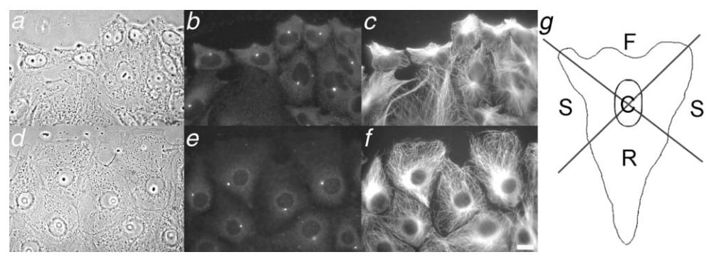
Fig.6 – Centrosome behavior in wound-edge CHO and PtK cells. Phase images (a and d) are shown, along with corresponding immunolocalization of γ-tubulin (b and e) and microtubules (c and f). Bar, 5 mm. (g) Diagram of cellular regions used to score centrosome position. Front, sides, rear and center regions are labeled F, S, R, and C, respectively. The wounds are toward the top of the figure. Image extracted from Yvon et al. 2002.
The cell polarization during migration, controlled by Rho GTPases, was assessed using an in vitro wound [17]. The study demonstrated that the Rho GTPase Cdc42 is essential to keep cell polarity around a wound edge.
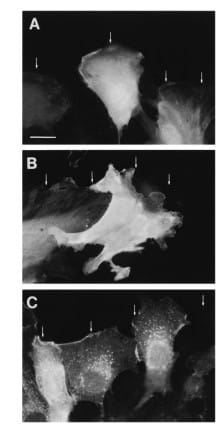
Fig.7 – Cdc42 is required for morphological polarization in leading edge cells. Rat embryo fibroblasts injected with a control fluorescent molecule (A) or with V12Cdc42 (C) correctly polarize at the wound edge, whereas cells injected with an inhibitor of Cdc42 activity (B) do not. Image extracted from Nobes et al. 1999.
Cell reorganization at the edge of a wound can be triggered by the forces that the cells sense, or rather the gradient of forces as the cells go from a continuous sheet to an abrupt stop. In epithelial cells, the tension felt by the nucleus was demonstrated to increase at the edge of a wounded sheet, reflecting the mechanisms that may trigger cell reorientation and polarization [18].
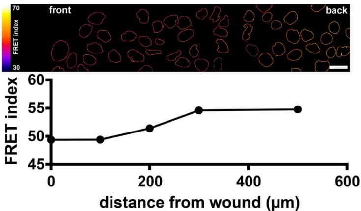
Fig.8 – The FRET index of artificial nuclear envelope protein constructs decreases at the edge of the wound (front, at left), indicating an increase in tension in nuclear envelope proteins of MDCK cells at the edge of the wound. Image extracted from Déjardin et al. 2020.
The wound healing assay can be an effective tool to study the effect of specific drugs on cell migration into the wound. Many examples exist in the literature, with researchers using the wound healing assay to assess the effect of medicines on migration of cells into wounds, for example to study the invasion of cancer cells [19, 20]. As an example, Kauanova et al tested the effect of microtubule-inhibiting drugs on in vitro wound healing assays to determine their effects on the kinetics of wound healing [2].
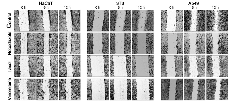
Fig.9 – Comparison of the time course of wound closure with the anti-microtubule drugs in a 12 h interval shows that the untreated cells HaCaT, 3T3, and A549 advance significantly into a wound, while the scratch area closure is slowed in the presence of nocodazole, Taxol, and vinorelbine. Image extracted from Kauanova et al. 2021.
The wound healing assay facilitates the acquisition of quantitative data related to cell migration in the healing process. Indeed, the well-defined cell-free gap mimicking the wound assures accurate and reproducible measurements of cell migration. Additionally, monitoring the wound closure over time using phase contrast microscopy or by using live cell imaging allows the analysis of migration parameters such as the wound area, the rate of cell migration and the wound closure percentage [21].
Thus, the wound healing assay represents a key tool for quantifying the healing process in vitro.
For example, the rate of wound closure can be assessed thanks to object detection by image segmentation. First, the original background of the image needs to be subtracted using ImageJ built-in “rolling ball” algorithm. Then, CellProfiler, a cell image analysis software, is used to calculate the area occupied by cells allowing easy quantification of the cell migration over time.
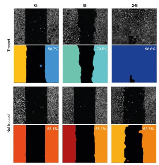
Fig.10 – Setting up the wound healing assay using a 4DCELL dynamic micropatterned coverslip. Images of the substrate were acquired at three timepoints: immediately after triggering cell migration with the reagent, 8 hours after and 24 hours after. A non-treated negative control is also provided.
Moreover, image J plugins can facilitate high throughput image analysis of in vitro wound healing images. The Wound Healing Size Tool, an ImageJ/Fiji plugin was designed for the quantification of wound area, wound coverage of total area, average wound width and width standard deviation [22].
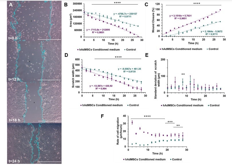
Fig. 11 – Wound healing size tool test comparing HaCaT exposed to hAdMSCs conditioned medium and HaCaT cultured only with basic medium as control of the assay. (A) Time lapse (0, 12, 18 and 24 hours. Scale bar = 27550 μm) images of wound healing closure in HaCaT exposed to hAdMSCs conditioned medium. (B) Scratch area in μm2. (C) Percentage of closure area. (D) Scratch width in μm. (E). Standard deviation of the scratch width in μm. (F) Rate of cell migration in μm/hour. Image extracted from Suarez-Arnedo et al. 2020.
Several products and techniques are available to perform in vitro wound healing. The traditional method is the scratch assay where a wound is created mechanically by applying pressure on a confluent cell monolayer. Tools like a pipette tip, a syringe, a toothpick or a cell scraper can be used for this purpose. However, these tools can damage the surface coating, potentially affecting cell migration and adhesion. The primary concern with this approach is the difficulty to obtain the same cell-free gap area between two experiments leading to a lack of reproducibility. Moreover, the poor precision of the utensil used makes it nearly impossible to obtain a well-defined and uniform wound. Thus, accurate monitoring and quantification of cell migration can become difficult to achieve [3].
To avoid these issues, substitutes to the scratch assay have been developed. The wound area can for instance be created with laser photoablation. This method is more precise than the manual one but can lead to unwanted cell death [23]. In their study, Wu et al. developed a wound healing assay based on ultraviolet light ablation [24]. With this method, wounds of different geometry and shapes can be produced. Nevertheless, the boundary of the wound is not well-defined with this method.
Another technique available involves creating a wound using a silicone insert [3]. While this device enhances precision and reproducibility, it is not entirely optimal, as the manual removal of the insert can result in non-uniform wound and variability in the results.
The wound healing assay developed by 4Dcell aims to answer all of the issues described above. This assay utilizes a micropatterning technique where the wound area is precisely printed using UV prior to cell seeding. The shape and dimension of the gap can consequently be customized to the user’s needs. The cells are first patterned in cell-adhesive regions and do not adhere outside of these regions. The surrounding anti-adhesive regions can be rendered adhesive by adding a single reagent onto the substrate. This induces cell migration into the newly adhesive regions making the micropattern dynamic.
In summary, the wound healing assay is a valuable in vitro tool to perform cell migration studies. The well-defined cell-free gap allows accurate quantification of cell migration and ensures experimental reproducibility. These assays have the potential of covering various physiological processes and pathologies related to cell migration. They reflect the behavior of cell sheets as well as the morphological aspects of cells during migration, which makes these tools multifunctional in their applications.
[1] Trepat X, Chen Z, Jacobson K. Cell migration. Compr Physiol. 2012 Oct;2(4):2369-92. doi: 10.1002/cphy.c110012. PMID: 23720251; PMCID: PMC4457291.
[2] Kauanova S, Urazbayev A, Vorobjev I. The Frequent Sampling of Wound Scratch Assay Reveals the « Opportunity » Window for Quantitative Evaluation of Cell Motility-Impeding Drugs. Front Cell Dev Biol. 2021 Mar 11;9:640972. doi: 10.3389/fcell.2021.640972. PMID: 33777948; PMCID: PMC7991799.
[3] Cappiello, F, Casciaro, B, Mangoni, ML. A Novel In Vitro Wound Healing Assay to Evaluate Cell Migration. J. Vis. Exp. (133), e56825, doi:10.3791/56825 (2018).
[4] Shi A, Li J, Qiu X, Sabbah M, Boroumand S, Huang TC, Zhao C, Terzic A, Behfar A, Moran SL. TGF-β loaded exosome enhances ischemic wound healing in vitro and in vivo. Theranostics. 2021 Apr 30;11(13):6616-6631. doi: 10.7150/thno.57701. PMID: 33995680; PMCID: PMC8120220.
[5] Zhang J, Chen C, Hu B, Niu X, Liu X, Zhang G, Zhang C, Li Q, Wang Y. Exosomes Derived from Human Endothelial Progenitor Cells Accelerate Cutaneous Wound Healing by Promoting Angiogenesis Through Erk1/2 Signaling. Int J Biol Sci. 2016 Nov 25;12(12):1472-1487. doi: 10.7150/ijbs.15514. PMID: 27994512; PMCID: PMC5166489.
[6] Lamalice L, Le Boeuf F, Huot J. Endothelial cell migration during angiogenesis. Circ Res. 2007 Mar 30;100(6):782-94. doi: 10.1161/01.RES.0000259593.07661.1e. PMID: 17395884.
[7] Beyer S, Koch M, Lee YH, Jung F, Blocki A. An In Vitro Model of Angiogenesis during Wound Healing Provides Insights into the Complex Role of Cells and Factors in the Inflammatory and Proliferation Phase. Int J Mol Sci. 2018 Sep 25;19(10):2913. doi: 10.3390/ijms19102913. PMID: 30257508; PMCID: PMC6213254.
[8] Hu Y, Rao SS, Wang ZX, Cao J, Tan YJ, Luo J, Li HM, Zhang WS, Chen CY, Xie H. Exosomes from human umbilical cord blood accelerate cutaneous wound healing through miR-21-3p-mediated promotion of angiogenesis and fibroblast function. Theranostics. 2018 Jan 1;8(1):169-184. doi: 10.7150/thno.21234. PMID: 29290800; PMCID: PMC5743467.
[9] Nareshkumar, R.N., Sulochana, K.N. & Coral, K. Inhibition of angiogenesis in endothelial cells by Human Lysyl oxidase propeptide. Sci Rep 8, 10426 (2018). https://doi.org/10.1038/s41598-018-28745-8
[10] Gerstberger S, Jiang Q, Ganesh K. Metastasis. Cell. 2023 Apr 13;186(8):1564-1579. doi: 10.1016/j.cell.2023.03.003. PMID: 37059065; PMCID: PMC10511214.
[11] Zhang, H., Zhu, C., He, Z. et al. LncRNA PSMB8-AS1 contributes to pancreatic cancer progression via modulating miR-382-3p/STAT1/PD-L1 axis. J Exp Clin Cancer Res 39, 179 (2020).
[12] Zhang M, Hong X, Ma N, Wei Z, Ci X, Zhang S. The promoting effect and mechanism of Nrf2 on cell metastasis in cervical cancer. J Transl Med. 2023 Jul 4;21(1):433. doi: 10.1186/s12967-023-04287-0. PMID: 37403143; PMCID: PMC10318801.
[13] Kurosaka S, Kashina A. Cell biology of embryonic migration. Birth Defects Res C Embryo Today. 2008 Jun;84(2):102-22. doi: 10.1002/bdrc.20125. PMID: 18546335; PMCID: PMC2542983.
[14] Szabó A, Mayor R. Mechanisms of Neural Crest Migration. Annu Rev Genet. 2018 Nov 23;52:43-63. doi: 10.1146/annurev-genet-120417-031559. PMID: 30476447.
[15]Alvizi, L., Nani, D., Brito, L.A. et al. Neural crest E-cadherin loss drives cleft lip/palate by epigenetic modulation via pro-inflammatory gene–environment interaction. Nat Commun 14, 2868 (2023). https://doi.org/10.1038/s41467-023-38526-1
[16] Yvon AM, Walker JW, Danowski B, Fagerstrom C, Khodjakov A, Wadsworth P. Centrosome reorientation in wound-edge cells is cell type specific. Mol Biol Cell. 2002 Jun;13(6):1871-80. doi: 10.1091/mbc.01-11-0539. PMID: 12058055; PMCID: PMC117610.
[17] Nobes CD, Hall A. Rho GTPases control polarity, protrusion, and adhesion during cell movement. J Cell Biol. 1999 Mar 22;144(6):1235-44. doi: 10.1083/jcb.144.6.1235. PMID: 10087266; PMCID: PMC2150589.
[18] Théophile Déjardin, Pietro Salvatore Carollo, François Sipieter, Patricia M. Davidson, Cynthia Seiler, Damien Cuvelier, Bruno Cadot, Cecile Sykes, Edgar R. Gomes, Nicolas Borghi; Nesprins are mechanotransducers that discriminate epithelial–mesenchymal transition programs. J Cell Biol 5 October 2020; 219 (10): e201908036.
[19] Li, H., Fan, Y., Yang, F., Zhao, L., & Cao, B. (2018). The coordinated effects of Apatinib and Tripterine on the proliferation, invasiveness and apoptosis of human hepatoma Hep3B cells. Oncology Letters, 16, 353-361.
[20] Lv, Y., Wang, W., Liu, Y., Yi, B., Chu, T., Feng, Z., … & Wang, Y. (2023). Platycodin D represses β-catenin to suppress metastasis of cetuximab-treated KRAS wild-type colorectal cancer cells. Clinical & Experimental Metastasis, 40(4), 339-356.
[21] Grada A, Otero-Vinas M, Prieto-Castrillo F, Obagi Z, Falanga V. Research Techniques Made Simple: Analysis of Collective Cell Migration Using the Wound Healing Assay. J Invest Dermatol. 2017 Feb;137(2):e11-e16. doi: 10.1016/j.jid.2016.11.020. PMID: 28110712.
[22] Suarez-Arnedo A, Torres Figueroa F, Clavijo C, Arbeláez P, Cruz JC, Muñoz-Camargo C (2020) An image J plugin for the high throughput image analysis of in vitro scratch wound healing assays. PLoS ONE 15(7): e0232565.
[23] Zordan MD, Mill CP, Riese DJ 2nd, Leary JF. A high throughput, interactive imaging, bright-field wound healing assay. Cytometry A. 2011 Mar;79(3):227-32. doi: 10.1002/cyto.a.21029. Epub 2011 Feb 9. PMID: 22045642; PMCID: PMC3306835.
[24] Wu SY, Sun YS, Cheng KC, Lo KY. A Wound-Healing Assay Based on Ultraviolet Light Ablation. SLAS Technol. 2017 Feb;22(1):36-43. doi: 10.1177/2211068216646741. Epub 2016 Jul 10. PMID: 27139694.
[25] Jonkman JE, Cathcart JA, Xu F, Bartolini ME, Amon JE, Stevens KM, Colarusso P. An introduction to the wound healing assay using live-cell microscopy. Cell Adh Migr. 2014;8(5):440-51. doi: 10.4161/cam.36224. PMID: 25482647; PMCID: PMC5154238.
[26] Vang Mouritzen M, Jenssen H. Optimized Scratch Assay for In Vitro Testing of Cell Migration with an Automated Optical Camera. J Vis Exp. 2018 Aug 8;(138):57691. doi: 10.3791/57691. PMID: 30148500; PMCID: PMC6126681.
[27] Kramer N, Walzl A, Unger C, Rosner M, Krupitza G, Hengstschläger M, Dolznig H. In vitro cell migration and invasion assays. Mutat Res. 2013 Jan-Mar;752(1):10-24. doi: 10.1016/j.mrrev.2012.08.001. Epub 2012 Aug 23. PMID: 22940039.